Endodontics

Endodontics, or root canal therapy, is that branch of dentistry that deals with the devitalisation of teeth.

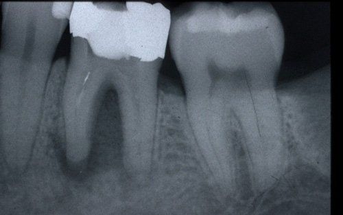
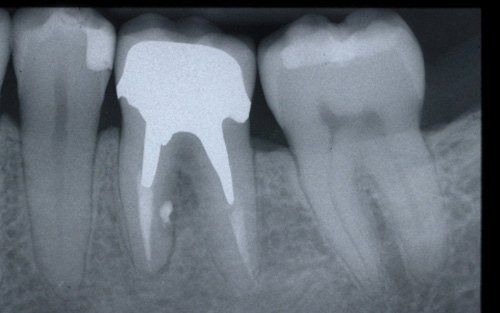
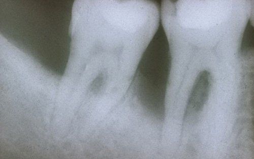
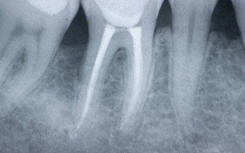
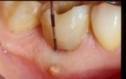
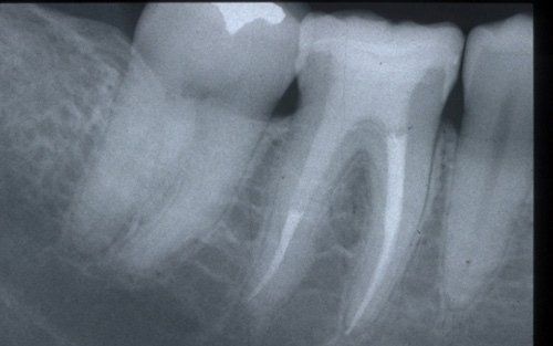
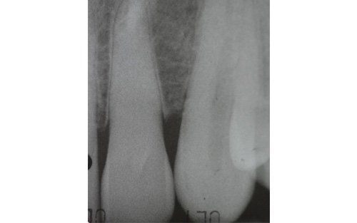
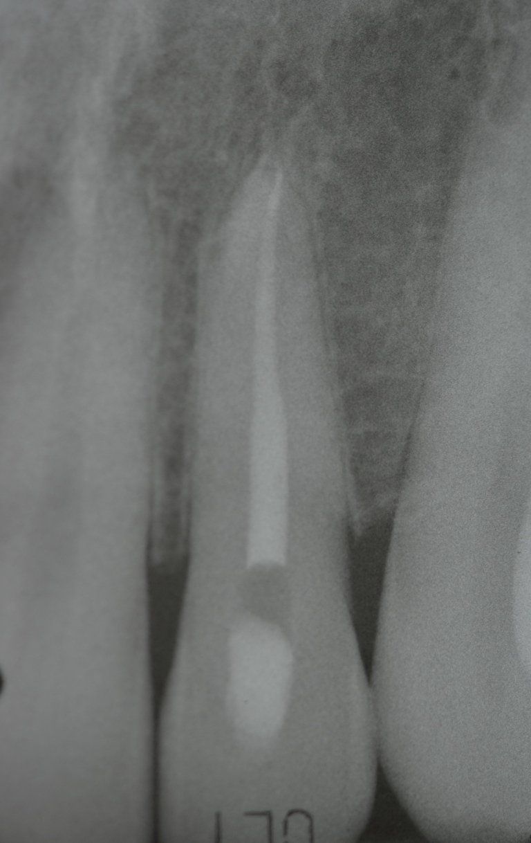
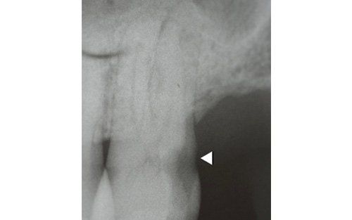
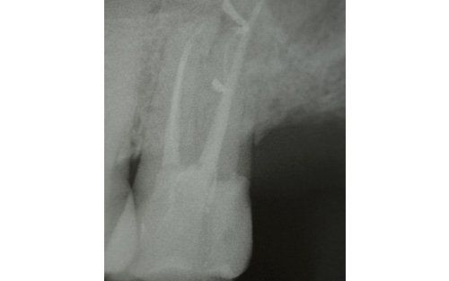
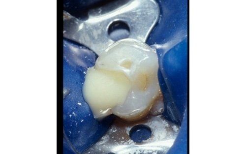
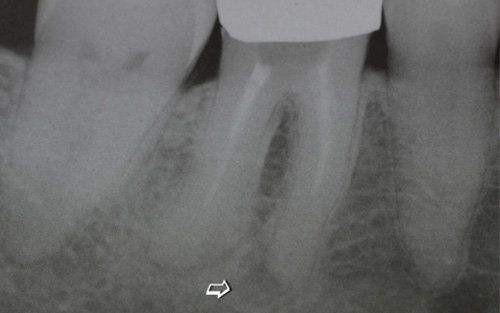
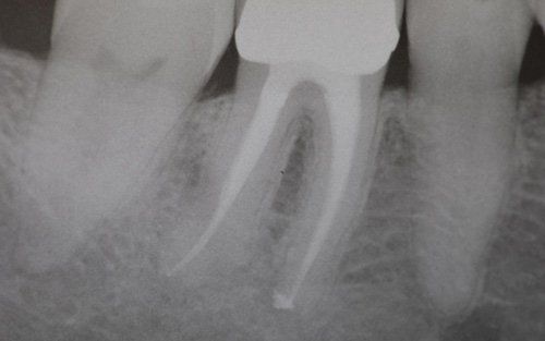
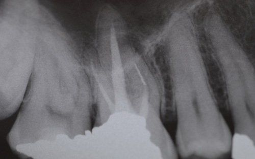
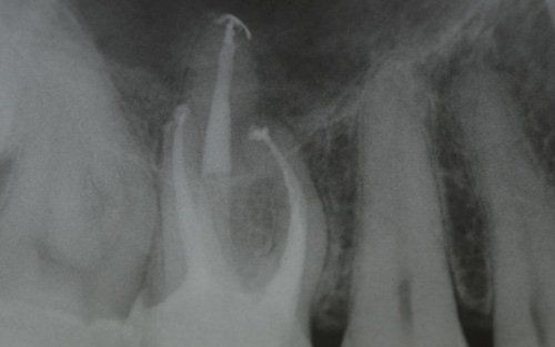
This treatment should be reserved for teeth that have irreversible pulp pathology (the nerve of the tooth) or an infection of the dental canals that has caused an abscess (acute apical periodontitis) or a granuloma (chronic apical periodontitis). While in the first two cases the tooth is very painful and the patient immediately seeks the dentist, the case of the chronic form is often silent, and therefore the patient may not notice anything for a medium to long period of time. In such conditions, it is only an X-ray image, taken during routine check-ups, that detects its presence. Root canal therapy can be completed by placing a latest-generation post inside the canal, as a means of retaining the material used for the reconstruction of the tooth. It will be up to the clinician to decide whether or not the restoration requires this additional anchoring system.

The endodontic treatment
Extensive destruction of the periradicular bone (the dark portion around the root). Also note the presence of a small fractured instrument in the mesial root. The tooth must be properly retreated.
The treated case. A radiographic check-up at 5 years after healing.
Extensive endo-periodontal lesion that extensively involved the periradicular bone of the elements of the lower right quadrant.
Tooth 46 after endodontic therapy and subsequent periodontal therapy. Healing is complete.
The cause of the fistula is a root perforation. The operator is using a periodontal probe to better understand the extent of the endodontic lesion.
Correctly devitalised and reconstructed tooth with the aid of a quartz fibre post, positioned in the distal root, to increase the retention surface of the restorative material.
The lateral incisor was correctly devitalised after difficult localisation of the calcified canal (previous image).
Note the extensive distal caries (indicated by the arrow). In the next image, we see the tooth correctly devitalised (note the presence of numerous lateral canals) and correctly reconstructed.
Pus discharge upon tooth opening. The patient, in these cases, experiences severe pain due to the pressure that the abscess material exerts on the surrounding tissues.
The arrow highlights a periapical granuloma (chronic apical periodontitis).
The tooth after endodontic therapy.
Upper molar with periapical granuloma on the mesio-buccal root: note the presence of a fractured instrument inside the root.
The tooth after retreatment is completed.
Share by: